


AI-enhanced MRI reveals cortical lesions in multiple sclerosis
A study reanalysed standard MRI scans from a large multiple sclerosis clinical trial using multicontrast processing and deep learning, improving detection of cortical lesions.
Source: AI-enhanced MRI reveals cortical lesions in multiple sclerosis (nature.com). Summary written with AI from the source and reviewed by rules; read the original.
What the research shows
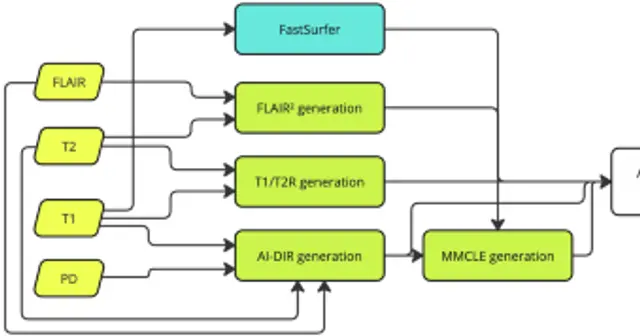
The study, published in the journal Communications Medicine, reanalysed standard MRI scans from a large multiple sclerosis clinical trial. The researchers combined multicontrast processing with deep learning, a type of artificial intelligence, to improve the detection of lesions in the cortex, the outer layer of the brain. According to the source, this approach improved detection of these lesions compared with the usual analysis of the same scans.
Cortical lesions are hard to see on routine scans, so detecting them has long been a challenge in multiple sclerosis research. Because this is a reanalysis of images already collected, the work shows an improvement in how these lesions can be seen, but it does not by itself show that they change the course of the disease or the response to treatment. Findings like this are usually expanded and confirmed by other groups before they influence clinical practice.
What it means
The finding suggests that computational techniques can make visible cortical lesions that might be missed on standard scans. This may help researchers track disease progression and the effect of treatments. It is still unclear how this improved detection will translate into care decisions for patients.
How to read it with care
This study does not replace a medical consultation. Anyone with multiple sclerosis or questions about scans and treatments should talk to their doctor or another qualified health professional before making any change.